

We are here to help you with your eye care needs. We want to let you know of all the services that are available at our office. We specialize in personalized service with a focused attention to detail.
In addition to complete eye examinations, digital imaging services, visual field testing, topography, pachymetry, Laser Vision co-management and contact lens services we are pleased to provide complete medical eye services.
Just an FYI, the following eye conditions can be treated here in our office:
✓ Eye Infections
✓ Eye Injuries
✓ Conjunctivitis
✓ Pink Eye
✓ Foreign Body Removal
✓ Eye Injuries
✓ Conjunctivitis
✓ Pink Eye
✓ Foreign Body Removal
✓ Eye Allergies
✓ Corneal Abrasions
✓ Dry Eyes
✓ Styes
✓ Lid Infections
✓ Corneal Abrasions
✓ Dry Eyes
✓ Styes
✓ Lid Infections
We are pleased to offer these special services in addition to our comprehensive eye care services. We are dedicated to providing each of our patients the finest eye care available anywhere. Our primary concern is to give you the very best of care.

-
About Dr. Sylvia
About Dr. Scott Sylvia
Scott Sylvia OD, MBA
Dr. Scott Sylvia did his undergraduate training at UMASS Dartmouth and received his Bachelor of Science in 1984 and his Doctorate in Optometry in 1986 from the New England College of Optometry in Boston. In 2000, he received his MBA with a concentration in Health Care administrative management and Marketing from Bryant College.
He worked with Charles Calenda MD in RI from 1986-1989, and 2010-2016, was a staff optometrist at Harvard Pilgrim Health Care in Rhode Island from 1989-1997. He opened his Cambridge office in Feb 1998 and joined Advanced Eyecare Associates in Providence, RI in 2016.
Dr. Sylvia performs primary eye care services and specializes in custom contact lenses including scleral lenses and hard to fit custom soft and gas permeable lenses. He is part of the Leadership Team from Vistakon / Johnson and Johnson, and has lectured and trained his peers in specialty contact lenses. He is also part of the Clinical Faculty from the New England College of Optometry and trains interns in his offices.
He is an affiliate doctor of the TLC Laser Eye centers and co-manages patients who undergo laser vision correction. He is a member of the American Optometric Association, the AOA Contact lens and Cornea Section, The Rhode Island Optometric Association, the Human Rights Campaign, Access Health MA, and the Greater Boston Business Council. He served as President of the Board of Directors of the Greater Boston Business Council. Currently he is the President of the Board of Directors of Access Health MA.

-
What Makes Us Different
What Makes Us Different?
Thorough, Unhurried Examinations
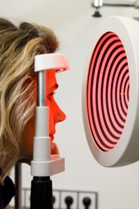
There are many approaches to performing eye exams. Some practices use technicians to perform tests and a quick visit with the doctor confirms the results. The Optometrists associated with Parrelli Optical® however prefer to take their time and get to know their patients and their visual needs. Our Doctors perform all the tests necessary for your eyes and then they discuss their findings with you and you decide on the course of treatment.
We Take the Time Your Eye Deserves
More than an eye test, your visit to one of the Independent Doctors of Optometry associated with Parrelli Optical® is a complete visual analysis and a comprehensive 15 point eye health assessment.
Our Doctors do much more than accurately determine your prescription. They will check your eye for common diseases like glaucoma or Age Related Macular Degeneration (ARMD) and problems like cataracts and presbyopia. In addition, many general health problems can be diagnosed early by assessing changes in the retina. High blood pressure, high cholesterol and diabetes first become visible when carefully looking at the back of the eye.
All testing is performed by qualified, independent Doctors of Optometry. -
Understanding Your Prescription
Understanding Your Prescription
Understanding the Numbers
Light bounces off everything we see. It then passes through an elaborate configuration of structures (parts of the eye) designed to bring that image to focus on the retina at the back of the eye.The Prescription Form
The Independent Doctors of Optometry associated with your eyecare-experts will record your vision correction on a form like this. Here’s what all those numbers mean:Your Prescription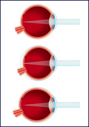
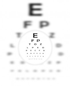
Emmetropia (top picture): When light entering the globe passes through the various parts of the eye and comes to focus right on the retina, the patient requires no vision correction and the form will be blank.
Hyperopia (middle picture): Usually termed “farsightedness.” Generally, this condition is noted by a lack of really clear vision at distance and near. However, the hyperopic patient, with some effort can often focus acceptably far away, in the distance. In this eyeball light focuses behind the retina. There is a lens (parts of the eye) within the eye that is used to add extra magnification for reading. Most farsighted people can use this lens to sharpen their distance vision. As this lens weakens, the doctor will prescribe magnifying lenses, written with a plus sign preceding the first number of the prescription (#1 on the form), to sharpen vision.
Myopia (bottom picture): Commonly called “nearsightedness,” this refractive error means that the patient can see well up close, within arm’s length. But, that vision is blurred in the distance. This is usually caused by the front of the eye being too strong (refractive myopia), or the eyeball being elongated (axial myopia). Both circumstances lead to light entering the eye coming to focus in front of the retina. A minifying lens, signified by a minus sign preceding the first number on the prescription pad (#1 on the form), focuses that light onto the retina, sharpening vision.Astigmatism: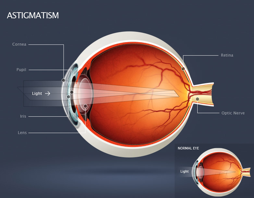
The clear window at the front of the eye is called the cornea. If it is the same curve in all directions, like a baseball, we call it spherical. If it is steep one meridian and flat in another, like a football, it is called astigmatic. Toric is Latin for “two curves.” We use a toric lens aligned with those two different meridians to sharpen vision. Astigmatism often compounds myopia or hyperopia, and in even modest amounts is designed into eyeglass lenses to sharpen vision. The amount of this correction is written in the second box of the prescription pad (#2 on the form), and its orientation is defined by the third box (#3 on the form).After being bent by the cornea light coming from objects closer than twenty feet away needs to be concentrated further in order to focus on the retina. The lens within the globe changes shape to provide this extra power. This lens is a multi layered structure very similar to an onion. Throughout life new layers are added. With time the center of the lens becomes further removed from its nutrients and it starts to harden. Ultimately, it will not flex enough to provide enough magnification for reading, and the patient will require reading glasses or multifocals. This additional power (ADD) is recorded under your near correction on the prescription pad (#5 on the form).
Prism:
Special light bending characteristics included for muscle imbalance. Represented by an amount and a direction entered into the final box on the prescription form (#4 on the form). -
Contact Lenses
Contact Lenses
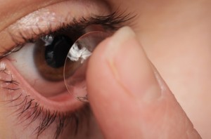
Anyone Can Wear Contact Lenses
Any prescription can be filled with contact lenses. Whether you are near sighted, far-sighted, have astigmatism or need a bifocals you can wear contacts. Contact lenses give you sharp vision, better peripheral field of view and improved depth perception.
There are literally an unlimited number of lens designs available today; soft and gas permeable materials, single vision and bifocal designs, spherical, aspheric and toric lenses. It takes a qualified professional to determine which lens will be best for you.-
Young Eyes Need Special Attention
- While spectacles are increasingly in vogue most young people tend not to want to wear eyeglasses. As a child comes of age in their adolescent years their self-image begins to form. Some research suggests that the inclusion of glasses in that image detracts from the development of self-confidence.
In the past the professional contact lens fitters have had concerns about fitting very young people to contact lenses. The failure to fully disinfect a lens once it has been opened and worn can lead to serious sight threatening complications. The complexity of the care regiment, for these re-useable designs only added to the potential for non compliance. In addition, these older lens designs slowed the flow of oxygen to the still growing eyeball and with little research to provide answers we always took the side of caution in waiting as long as possible to fit young eyes to contact lenses.
With the introduction of single use contact lenses, we are much more comfortable fitting younger eye with lenses. Single use contact lenses allow the wearer to open a fresh, clean and sterile contact lens each day. Now with more than 15 years of experience dispensing these modern lens materials and a wealth of international research, we are confident that young eyes can safely wear lenses for many years. -
Contacts in Adults
- New Wearers:

Contact lenses offer an unprecedented option for virtually every vision correction. New materials improve wearing comfort. Modern designs correct even the most complex prescriptions, even those including bifocals and astigmatism. Planned replacement designs minimize the care regime. Pop them in, see well, and be comfortable. See for yourself, call for your convenient appointment today.
Existing Wearers;
Years ago lens materials and designs were not as easy on your eyes as our newer innovations. If you have been wearing contacts for a long time and you may be experiencing periodic blurred vision, end of day dryness, inability to read up close, redness, irritation and discomfort. These are all signals that your lenses are insulting your eyes. Those little irritations go into a cumulative pool of reactions that ultimately leads to contact lens intolerance and an inability to continuing wearing contacts.
We do not fit contact lenses, we fit eyes.
For more than twenty five years we have been committed to studying, researching and introducing the newest, safest healthiest contact lens options available in the marketplace. We want you to be able to wear your lenses where and when you wish. We go to great lengths to assure that you can have a lifetime of comfortable lens wear. -
A Contact Lens is a Medical Device
- The cornea is the clear window on the front of your eye. It comprises a vast majority of the transparent tissue in the human body. It is a remarkable structure; five individual layers of living tissue dependent upon a myriad of mechanisms to keep its clarity while providing you with sharp vision. When you place a contact lens on this delicate structure you disrupt its intricate balance. For every individual cornea there is a well fitting contact lens.
It is the job of your eyecare professional to assure that tiny piece of plastic floats gently on the tears preserving the integrity of the tissue beneath while providing clear sharp vision. Not just today but as you wear those contact lenses day in and day out over a lifetime. Because your vision is just important to you now as it will be twenty, thirty or even fifty years from now.
We have access to designs from every major manufacturer. We do not push one model. Through an extensive evaluation of your eyes we will determine the best contact lens for you. The reason that there are a virtually unlimited number of contact lens designs is that every eye is distinctly different. More distinct than a fingerprint, each eyeball requires something unique to assure the comfort and vision of a contact lens. We always think about what is best for you.
We have established relationships with designers and manufacturers of soft, gas permeable, rigid and hybrid contact lenses. We have the skill and experience to fit each of these specialty designs to assure your comfortable wear, good vision and ongoing eye health.
We use the latest innovations in equipment to measure your eyes to guaranty your comfort, vision and continued eye health. -
Complex Prescriptions Require Specialize Fitting
- The cornea is the clear window on the front of your eye. It comprises a vast majority of the transparent tissue in the human body. It is a remarkable structure; five individual layers of living tissue dependent upon a myriad of mechanisms to keep its clarity while providing you with sharp vision. When you place a contact lens on this delicate structure you disrupt its intricate balance. For every individual cornea there is a well fitting contact lens.
More complex prescriptions typically require more complex lenses. If you a very high degree of astigmatism, nearsightedness, farsightedness , need a bifocal or have very dry eyes you should consider rigid gas permeable lens designs. These lenses are somewhat firmer than soft lenses. But, that allows them to provide a new soother surface for the cornea resulting in much sharper vision.
If you have had an injury, suffer from a disease or have had surgery on your eyes the surface of your cornea may be irregular. A soft lens drapes over the cornea and that irregularity will show through leaving you with unacceptable vision. That same soft lens may slow the flow of oxygen, the main source of nutrition for corneal tissue, interrupting the healing or compromising the health of your cornea.
Rigid gas permeable contact lenses can be just as comfortable to wear as soft contact lenses. But, they need to be designed by an experience rigid contact lens fitter to assure your comfort and the health of your eyes.
We specialize in the fitting of contact lenses for corneal ectasia, keratoconus, pellicid marginal dystrophy, post graft corneal transplant, strong prescriptions and irregular corneas. -
What You Need to Know about Contacts
- Contact Lenses come in many forms and perform many functions.
Soft Contact Lenses:
Soft contact lenses are soft, supple and they even fold in half. But, it is their size that makes them so tolerable. They are large enough to fit well under the eyelids and are therefore remarkably comfortable, even if you have never worn them before. These designs come in a wide range of replacement schedules.
Colored Contact Lenses:
These lenses come in a wide array of colors. They can enhance your natural eye color or change that color completely. Modern technologies give you a natural look in the eye color that you have always wanted.
Theatrical Lenses:
For those interested in a wide variety of contact lens designs from extreme vivid colors to wild eye designs, we fit an extensive collection of theatrical lenses for your performance on the stage or in your life!
Lenses for Astigmatism:
A contact lens rests on the clear window in front of the colored part of your eye. Rarely is this tissue the same shape in all directions. Most eyes have a small amount of irregularity called astigmatism. Most eyes need a correction for a refractive error (see your prescription) that can be provided by a standard or spherical contact lens. If you have a greater amount of astigmatism your eyecare professional will fit you to a “toric” lens.
One of the reasons that a soft lens is so comfortable is that it drapes over your cornea. But, this allows an underlying irregular shape to transfer through the lens. A spherical contact lens will allow you to see better than with no correction, you will not see as well as you do with your glasses. “Toric” is Latin for two curves, and just like your spectacles, a toric lens has two curves cut on the back of it which align to the different curves on the front of your eye. These lenses provide great comfort and great vision.
Multifocal Contact Lenses:
Single vision lenses let you see in the distance. When you also need help with reading, we can fit you to a bifocal contact lens. Spectacle bifocals have the distance correction on the top of the lens and the extra power for reading at the bottom. We have never been able to create a really effective contact lens that has allowed us to keep the reading segment in the right position. So all bifocal contact lenses use some combination of concentric rings. This allows you to see at distance and at near. We offer experienced contact lenses fitters who will use the latest innovations to provide you with good vision at distance, near and everywhere in between.
Rigid Gas Permeable Lenses:
While once reserved for the more complex prescriptions, high degrees of myopia or large amounts of astigmatism, rigid lenses are once again, increasingly popular. Originally we made rigid lenses from a plexiglass like product, the material used for storm doors. This plastic provided great clarity of vision, but did not allow oxygen to pass through to and provide nutrition for the cornea. As a result, we had to make the lens very flat so that it would rock and draw oxygenated tear film under the lens. This meant that the bottom edge of the lens literally stuck out and bumped the lid as the wearer blinked.
Modern gas permeable materials allow a high volume of oxygen to pass right through the lens. And, modern manufacturing technologies allow us to use complex designs that mimic the shape of the cornea, vastly improving wearer comfort. -
Offering the Greatest Comfort
- Soft contact lenses are in fact soft. They are very supple; they can even be folded or turned inside out. When a contact lens is on your eye it can be more than half tear film. But, it is the size of the lens that makes it so very comfortable. The lens fits well underneath your lids, limiting the interaction between the lens and the edge of your lid providing astonishing comfort, even if you have never worn a lens before.

New wearers need to build their tolerance to contact lenses by slowly increasing their daily wear times over their first week of wear. While, your Parrelli Optical® contact lens specialist can see adaptive symptoms on the front of your eye through your first weeks, most patients have no real symptoms after the first few hours with lenses on.
Replacement Schedules:
Many years ago we gave patients a single pair of lenses and a rather complex care kit and said “Try to make these last six to twelve months.” Soft lenses are about 50% tear film when they are on your eye. As the tear passed through the lens it deposited organic substances like calcium, lipid and mucus on the contact. Environmental contaminants also collected on the lens surface. As a result by the end of the 1980’s we were sending patients home with a burdensome chemistry kit to care for their lenses. As you might imagine, compliance was an issue and we began to see a wide variety of complications directly attributable to dirty lenses.
In 1988 manufacturing engineers studied the problem and created new ways to fabricate contact lenses. The results are better quality products in a wider array of designs that can be affordably replaced on some kind of a pre-planned schedule.
One Day: These lenses are opened from a sterile package, worn and thrown away after one use. There is no care regime required. One day lenses are the healthiest and safest way to wear contacts. They are exceptionally convenient and are recommended for young eyes.
Two week: This lens is designed to be used 14 times or be open for 30 days. If you are wearing lenses every day, you would replace them after two weeks. Care of these lenses requires a multi-purpose solution. After removal, the lenses are rubbed, rinsed and stored, all with one product. Until recently this was the most popular modality.
Monthly: Increasingly popular monthly designs offer a greater variety of correction options (astigmatism and bifocal designs in particular). Because we do so little on a two week schedule, the increase in popularity of this lens can be traced to the simple fact that it is easier to remember to replace your contacts once a month than to remember to do it every two weeks. The longer replacement schedule requires a more aggressive care system. Each day after removal, the lens is rinsed and then stored for six hours in a more effective cleaning and disinfecting agent. This is the modern paradigm, most manufacturer’s newest designs are in the monthly format.
Quarterly: Highly specialized designs for unique prescriptions may require custom designed lenses that are replaced every two to three months. These require a significant increase in daily maintenance to be safely cleaned and disinfected. -
Providing Superior Vision
- Providing Superior Vision:
Rigid or gas permeable lenses are firm. Rather than draping over the cornea they float on top of that delicate tissue. Each lens is in essence two lenses. The first lens is the contact itself which forms a new, smooth surface for the front of eye. The second lens is the layer of tear between the back of the contact and the front of the cornea.
Damaged Corneas and Eye Disease:
Your Parrelli Optical® skilled, professional contact lens fitter can use this unique lens design to create a tear layer that will neutralize any corneal irregularity that might be found in a damaged or diseased tissue, including keratoconus, pellucid marginal degeneration of corneal transplants.
Complex Prescriptions:
Modern, computer generated manufacturing technologies allow us to fabricate far more intricate designs than ever before. At Parrelli Optical® we use our expertise to design lenses for complex prescriptions like astigmatism, bifocals and multifocals. Each of our gas permeable lenses is individually designed and custom fabricated for your eye affording almost anyone comfortable wear and great vision.
Dry Eye:
Modern soft lenses are generally 50% tear film when they are on the eye. That means you need to produce enough tear to keep that lens hydrated, comfortable and functioning well. A rigid lens material is less than five percent water, so that you only need enough tear to float that lens on your eye. Many dry eye patients find gas permeable contact lenses quite comfortable.
Rigid Gas Permeable contact lenses remain the only responsible annual replacement contact lenses. A daily cleaner and separate storage solution for safe disinfection and healthy lens wear are indicated on a daily basis.
Gas permeable contact lenses require an extra level of professional expertise to design and build. Our eyecare professional has the experience to improve your vision and your comfort.
Exciting New Lens Designs
Gas permeable contacts are firm and yield exception sharpness of vision. They can correct a wider range of vision problems. The firm lens floats on the front of the eye providing new ocular surface for irregular, diseased or surgically altered corneas. These lenses also provide much more oxygen to the delicate cornea that other materials.
Some eyes, due to genetics, damage or injury are hyper-sensitive to contact lenses. A hybrid contact lens provides the best answer for these patients. Hybrid lenses are made from two different plastics. The center portion of a hybrid contact lens is a gas permeable lens that provides high definition vision. The periphery of the hybrid contact is soft. This outer ring is supple and it slides under your eyelids to provide exceptional wearing comfort.
Benefits:
Hybrid contact lenses can offer you the best of both worlds. Your brain receives the high-definition visual acuity and your eye receives the extra oxygen associated with gas permeable lenses, but more importantly, you also get exceptional comfort delivered by soft contact lenses. Hybrid contact lenses are indicated for keratoconus, corneal ectasia, problems reading, high amounts of astigmatism and for the hypertensive eye that is intolerant of other designs. -
Exciting New Lens Designs
- Gas permeable contacts are firm and yield exception sharpness of vision. They can correct a wider range of vision problems. The firm lens floats on the front of the eye providing new ocular surface for irregular, diseased or surgically altered corneas. These lenses also provide much more oxygen to the delicate cornea that other materials.
Some eyes, due to genetics, damage or injury are hyper-sensitive to contact lenses. A hybrid contact lens provides the best answer for these patients. Hybrid lenses are made from two different plastics. The center portion of a hybrid contact lens is a gas permeable lens that provides high definition vision. The periphery of the hybrid contact is soft. This outer ring is supple and it slides under your eyelids to provide exceptional wearing comfort.
Benefits:
Hybrid contact lenses can offer you the best of both worlds. Your brain receives the high-definition visual acuity and your eye receives the extra oxygen associated with gas permeable lenses, but more importantly, you also get exceptional comfort delivered by soft contact lenses. Hybrid contact lenses are indicated for keratoconus, corneal ectasia, problems reading, high amounts of astigmatism and for the hypertensive eye that is intolerant of other designs.
-
-
Anatonomy of the Eye
We Understand Eyeballs
The human eye is a miraculous structure, a one inch globe that focuses the entire world on tissue the size of a postage stamp. For you to see clearly, incoming light must come to a precise focus on the retina at the back of the eye. The entire structure and all its components are dedicated to that end.Parts of the eye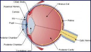
In this cross section of the human eyeball, we can trace the beginnings of vision. Light enters the globe through the cornea (far left). This tissue is clear and normally has no blood vessels. It receives all its nutrition from oxygen that it can derive from the environment and from the tear film that bathes it. The cornea contributes about two thirds of the focusing power of the eye. The tear film itself forms a smooth refractive surface bending light uniformly.
Next is the anterior segment. This area is divided into the anterior chamber, the space in front of the iris and posterior chamber behind the iris. This space is filled by a liquid, called the aqueous humor. This fluid is produced in the cilliary body, a radial structure that supports the iris.
This incoming light then encounters the iris. This reactive diaphragm varies in color from blue to green and most commonly to brown. In the center of this structure is a hole, called the pupil. The iris adjusts the pupil’s diameter in reaction to the intensity of incoming light. The brighter the light, the smaller the hole.
Aqueous humor is constantly produced behind the iris and flows across the surface of the crystalline lens through the pupil and exits via a sieve-like structure that surrounds the base of the iris called the trabecular meshwork located in the angle between the back of the cornea and the front of the iris.
Incoming light then encounters the crystalline lens. The lens is also clear and devoid of blood vessels. The constant flow of aqueous across its surface nourishes this tissue.
This structure has a natural tendency to bulge, to become thicker at its center. The lens is suspended in a capsule. In a straight gaze it contributes an additional one third of the eye’s focusing power. When you are looking at a distance, it is held compactly by the surrounding ciliary process, and not allowed to enlarge. When the ciliary muscle relaxes, the lens changes shape adding more magnification for reading. This occurs naturally as your eyes converge to focus on objects closer than twenty feet away.
Light then passes through the posterior cavity. This part is filled with the vitreous gel, a thick gelatinous substance, that does not regenerate with time. This supports the globe and holds the retina in place.
Finally, in an eye needing no vision correction, the light comes to a sharp focus on the retina. Covering the inside of the sphere, and extending in all directions all the way forward to its attachment at the ora serrata surrounding the base of the ciliary body, the retina is a vast network of light receptors. This is where vision begins.The retina uses a rich vascular network to fuel an electro-chemical reaction that turns light into sight. Light’s energy is translated into an electrical impulse by a chemical reaction that takes place at its receptors. The peripheral portion of this structure is composed of receptors known as rods. They are very sensitive to light and movement but only translate signals in black and white. The macula is a 3 to 5 mm oval, surrounding the fovea at the approximate center of the retina. This spot has the highest concentration of receptors called cones and is responsible for color perception and the sharpest vision. Retinal receptors are wired directly to the brain via nerve fibers.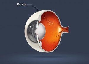
Millions of these nerve fibers gather to form the optic nerve, which exits through the only opening in the globe, and carries the electrical impulses generated by the retinal receptors to the visual cortex at the back of the brain where vision occurs.-
Eye Diseases inside of the Eye
- Eyes Rarely Hurt When There Is Something Wrong
There are many disorders of the eye. The following information briefly discusses the causes and treatments for the many of the more common inflammations, infections, effects of aging and diseases that we see regularly in our practice. The more you know about your eye’s health the more prepared you can be to deal with complications. These discussions are not intended to be used to diagnose your eye health symptoms.
Inside of the Eye
Sight Threatening Complications
Light bounces off of everything. That light is then gathered by the cornea, the clear front window of the eye. Pictures from your world then pass through the pupil, a hole in the center of the colored, circular part of your eye called the iris. Its path is then refined by the crystalline lens. This lens changes shape to add magnification to the eye when you focus on something closer than twenty feet away. Finally, the image from your world is focused on the retina where it is turned into electrical impulses which pass to the brain where they are interpreted as vision.
This is an obvious simplification of a complex interaction between many components that result in the miracle of sight. The slightest disruption to the harmony between the orchestrated interaction of all these components can have a profound impact on your ability to appreciate the world around you. What follows are explanations of what can impact the quality of your sight inside your eye.Glaucoma: Increased Eye Pressure and Its Effect on Vision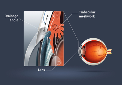
The cornea is the clear window at the front of your eye (parts of the eye). Directly behind it is the first section of the globe called the anterior segment. This compartment is divided into two chambers separated by the iris and filled by a liquid called the aqueous humor. Aqueous is constantly produced in the ciliary body, a radial structure in the posterior (back) chamber at the base of the iris. It then nourishes the crystalline lens as it flows across it, and exits through the pupil into the anterior (front) chamber.
Increased intraocular pressure can be caused by an increase in output or a decrease in the outflow of aqueous humor.
This fluid is then filtered through the trabecular meshwork and drains from the eye through a radial canal located in the angle formed by the front of the iris and the back of the cornea. With age, this angle narrows or the meshwork gets clogged with debris, slowing the outflow of aqueous. This triggers an increase in the pressure within the globe. Increasing intraocular pressure pushes the optic nerve fibers against the sides of the hole through which it exits the eyeball.
Glaucoma slowly affects the peripheral vision. By the time the patient realizes it this vision is permanently lost.
Ultimately, this leads to a predictable pattern of peripheral vision loss. The patient is asymptomatic and can experience a significant and permanent loss of vision before they become aware of this condition.Cataracts: Sharper Vision the Next Day
What Is A Cataract?
Within the anterior chamber there is a lens that changes shape to add magnification to see things up close. This crystalline lens is structured like an onion, having a central nucleus that is surrounded by layers, and the whole body is held within a capsule. The capsule is suspended by zonules, that on straight gaze are connected to a radial muscle that pulls tightly compressing the lens. In a process known as accommodation, that muscle relaxes, the capsule slackens, and the lens naturally bulges, increasing magnification, allowing you to read comfortably.
With A Cataract, The Lens Within The Eye Becomes Cloudy
Throughout a lifetime, new layers are generated and added to the outside of the lens. As the center becomes more separated from its nutrients by these layers, it begins to harden and discolor as a cataract forms. This discoloration leads to cloudiness and eventually to an opacity. It is then considered a cataract. A cataract only becomes a surgical issue when it interferes with clear vision. When a patient becomes insecure with their vision, the lens can come out. Surgical intervention is the only effective method to treat cataracts. The opacified lens is removed and a plastic intra-ocular lens (IOL) is put in its place. This is one of the most common surgical procedures performed in this country. It incorporates a local anesthesia, and the patient usually returns home on the same day with functional vision with a full post-operative recovery within a few months.
With cataract surgery the natural lens is removed and replaced with an implant restoring vision miraculously, literally overnight.Floaters: Annoying Visual Distractions
Flashers and Floaters
Floaters
The vitreous is a clear gel-like substance that fills the back of the eye. It gives the globe it’s shape and holds the retina in place. It is not replenished over time and it does degenerate. With time the vitreous body shrinks and pulls away from the retina. This can release cells that float across the visual pathway, casting shadows in the field of view. Floaters present as spots, dots, lines circles or cobwebs that float across your line of sight. They are frequently visible when looking at a plain background such as a blank wall or a blue sky. These floaters represent benign changes and are typically age related with most complaints starting around age forty. They are usually of little importance. But a sudden onset or increase in intensity of floaters should be evaluated by your eyecare professional.
Flashers (flashing)
The retina is a thin , transparent tissue that lines the back of the eyeball. It uses sophisticated photoreceptors that gather light and through electro-chemical reactions, turn that light into vision. A tearing of this tissue or a separation of the retina from the back of the eye can result in flashes as if a light where flashing in your peripheral field of view. This can be followed by a blocking of your peripheral vision as if a shade where being drawn across part of your field of view. This is typically associated with a detachment and a folding of the retina and requires immediate attention to avoid permanent vision loss.Macular Degeneration: Blurred Central Vision
Macular Degeneration or Age Related Macular Degeneration (ARMD)
The macula is a 3 to 5 millimeter oval surrounding the fovea, which is located at the approximate center of the retina (parts of the eye). It has the highest concentration of retinal cones. These sensitive receptors relate colors and are responsible for the perception of fine detail, like face recognition and reading, it is the only portion of the retina that can achieve 20/20 vision. In age related macular degeneration, there is a breakdown in the delivery of nutrients to this region. This results in a thinning of the tissue. In addition, with time, debris deposits begin to appear in this region playing a part in macular degeneration. Each of these contributes to, and both in combination cause, a dimming or distortion to central vision.
Dry Age Related Macular Degeneration (ARMD):
Pigmented spots called drusen, thought to be debris from deteriorating tissue, begin to appear in macular degeneration.
This “dry” form of Age Related Macular Degeneration (ARMD) leads to mild vision problems and may leave the patient more susceptible to the “wet” form of this disease.
Wet Age Related Macular Degeneration (ARMD):
New blood vessels leak fluid killing light sensitive receptors.
The wet form of age related macular degeneration (ARMD) is a severe disorder and warrants immediate attention. New blood vessels growing under the fovea leak fluid. This causes the light sensitive cells near them to wane, resulting in a dramatic loss of central vision. While the peripheral field of view is impacted, the loss of central visual acuity has a tremendous impact on a patient’s motility.Posterior Uveitis: Inflammations of the Eyeball
“itis” means ‘to be inflammed’. ‘itis’ is often added to end of the name of a part of the body to describe an inflammatory condition of that part.
Keratitis
The corneal is the clear window at the front of your eye. Inflammation of this tissue is call keratitis. A superficial case of keratitis can be treated and will heal with minimal side effects. As a general rule the deeper the pathenogen penetrates the corneal tissue the more painful and severe the reaction will become. When this inflammation reaches the deeper layers of the cornea, treatment becomes more aggressive to minimize the resulting scarring. Scarring in the clear corneal tissue will result in some degree of vision loss.
Inflammation of the cornea can be viral, fungal or bacterial in nature. Over the counter medicines are ineffective and can lead to severe damage to your eye. Your doctor will assess your condition and prescribe an appropriate medication.
A much more serious form of the inflammation is ulcerative keratitis, in which an ulcer forms in the clear tissue of the cornea. Here the offending agent, again viral, fungal or bacterial, penetrates the outside layer of the tissue attacking the stroma in the deeper recesses of the cornea. This is an extremely painful condition that can lead to blindness. Ulcerative keratitis requires immediate medical attention. Your physician will prescribe an aggressive application of a condition-specific medication and monitor your response very closely.
Even with aggressive treatments, more intense inflammations can lead to varying degrees of scar tissue formation. This results in a loss of eyesight. In more dramatic presentations, after the resolution of the keratitis a corneal transplant may be necessary to restore some useful vision.
Piscleristis The tissue connecting the sclera, the white outside layer of the eye, to the conjunctiva, its overlying mucous membrane, is called the episclera. Episcleritis, an inflammation of this tissue is mild and rarely triggers the more severe scleritis. Scleritis The sclera is the white, tough fibrous membrane forming the outside tunic of the eye. When this tissue becomes inflamed it is called scleritis. This complication is often associated with systemic diseases, most commonly rheumatoid arthritis. It is also associated with disorders related to menstruation and is therefore more common in women, usually presenting after the age of forty. There are three levels of intensity associated with the presentation of scleritis. The most common is diffuse scleritis. In nodular scleritis the symptoms are more intense. With necrotizing scleritis being the most severe.Your physician will determine the most effect course of treatment. Anti-inflamatories, steroids, antibiotics and surgery for more dramatic cases are options available for treatment. Uveitis The uvea is the middle layer of the eye. It consists of the ciliary body which holds the crystalline lens behind the iris, the chroid which is the rich vascular network that nourishes the retina, and the iris which is the colored part of your eye that controls the amount of light entering the globe by adjusting the size of the pupil. Uveitis is a painful inflammation of some part of the Uvea. Uveitis is a general diagnosis used to define an inflammation within the eye. It can be further classified as front (anterior), middle (intermediate) or back (posterior). This inflammation can present in a single incident or in chronic reoccurring episodes. Steroids are used to quiet the body’s inflammatory response and are the first treatment choice for uveitis that is not bacterial in nature. The prognosis is usually good for patients receiving prompt diagnosis and treatment. Iritis The iris is the colored part of your eye, just behind the clear cornea. The hole in the center of this structure is the pupil. The iris regulated the amount of light entering the back of the eye. Inflammation of the iris is called iritis. Infections and disease are common triggering agents. But, many times no cause for the inflammation can be identified. Iritis can be acute or chronic. Acute iritis presents rapidly, can clear on its own or may require medications to quiet the response. Acute iritis rarely causes visual impairment. Chronic Iritis which will reoccur unexpectedly over many years is much more difficult to treat. Chronic iritis typically does not respond to medications. Information provided in the Website is intended to aid in your understanding of the eye and its operation; it will not replace your physician. Have you eyes examined annually.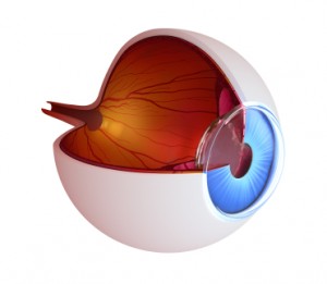 Diabetic Retinopathy: A Serious Threat to Vision
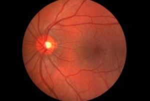
Diabetes can accelerate cataract (cataract) formation and induce glaucoma (glaucoma). More commonly the diabetic patient is susceptible to a series of changes in the back to the eye (retina) called diabetic retinopathy.
Vision comes from light entering the eye and focusing on the very sensitive receptors of the retina that lines the back of the globe. Detail driven central vision is provided by a concentration of receptors in the macula, located at the center of the retina. This tissue is supported by a rich vascular network that nourishes this tissue. Changes in the retina can be triggered by diabetes.
Non-proliferative Diabetic Retinopathy (Background Retinopathy):
Weakened blood vessels in the region of the macula can leak fluids causing the light receptors to be permanently damaged, causing central vision to deteriorate, in the early stages of diabetic retinopathy.
Proliferative Diabetic Retinopathy (Proliferation Retinopathy):
Delicate new blood vessels are formed. These fragile new vessels grow on the face of the retina and can rupture easily. These microscopic or micro-aneurysms bleed into the vitreous . The leaking blood creates a cloud that obscures the retinal receptors, blurring or blocking vision. Scar tissue can form at the site of the leak tightening and pulling the retina inducing detachments leading to severe vision loss.
Treatment for Diabetic Retinopathy:
The treatment of choice is photocoagulation, where small bursts of intense heat from a laser, focused on the leaking vessels, stops the bleeding. The resultant scars reduce abnormal vessel growth and help to “spot weld” the retina to the globe.
Early detection, exercise, controlling blood sugar levels and some preventative laser therapies can preserve your vision. See your eyecare practitioner annually. -
Eye Disease in Front of the Eye
- Many Problems Start Here
The eyeball is the only part of the human central nervous system that is exposed to the world. This delicate light gathering organ is responsible for upwards of 85% of the information provided to your brains. Words, pictures, scenes and vistas are all collected by the eye and sent to the brain to be processed into what we know as sight. The lids, lashes, tears, conjunctiva and the cornea all need to be in near perfect balance for you to see the world around you.
What follows are some of the more common complications that impact the front of the eye.Blepharitis: Red Irritated Lids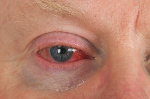
Blepharitis is an inflammation of the eyelids. Some people are more prone to blepharitis. Individuals of Celtic descent, light complexion, light hair and or light eyes are at greater risk. Their symptoms include red, swollen or crusty eyelids.
The tear film is made up of three layers. The inside layer is mucous that holds the middle aqueous, or watery layer, on the front of the eye. The outside layer is an oily film that is secreted by meibomian glands which empty along the edge of the lids just behind the lashes.
When the middle aqueous or watery layer of the tear film lessens, the oil from the meibomian glands is not diluted and it starts to collect at the base of the lashes. Blepharitis occurs when skin cells that slough off and would normally be carried away when you wash can now be trapped in this oily build up. These dead cells then become a source of bacteria which can cause infections within the lid or on the front of the eye. Blepharitis is of particular concern to contact lens wearers.
Careful attention to lid hygiene, particularly in times of season change can prevent blepharitis. When you wash your face, use a face cloth to gently rub along the edges of the closed eye to prevent the oily build up. Frequent warm compresses can also open the glands and purge them of debris. Ask the eyecare professionals at Parrelli Optical® about medicated scrubs to treat blepharitis.Red Eye, Conjunctivitis and Subconjunctival Bleed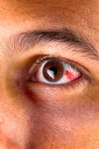
The eyeball has a limited number of ways to react to insult, the most common is redness. There is a thin mucus membrane (parts of the eye) that lines the inside of the eyelid and folds over to cover the outside of the eyeball. Inflammation or irritation of this tissue can lead to red eyes.
Subconjunctival Bleed: There is a vast network of blood vessels just below the surface of the white part of the eye. Even a single drop of blood leaking from the tip of one of these delicate vessels can lead to significant redness in the eye. Eye rubbing, coughing, sneezing and even the pressure of the finger tips pinching to remove a contact lens can result in this form of red eye. This can also be an indication of high blood pressure of other systemic disease. While in most cases the red eye will clear over several days you should always consult your eyecare practitioner for treatment
Bloodshot Eyes: The eye can become very dry due to systemic problems (see dry eye) or in reaction to transient environment conditions, medications, drug or alcohol use. In response to dry eyes the blood vessels just beneath the sclera (parts of the eye) fill with blood and become closer to the surface and cause red eye. Generally, drops designed to get rid of the this form of red eye, treat the symptoms by constricting those blood vessels, as these drops wear off the blood rushes back, enlarging the vessels further, requiring continuous use. Rewetting drops and whole body hydration treat the cause and resolve the red eye.
Conjunctivitis: An inflammation of the conjunctiva can lead to red eyes and has several causes.
Bacterial Conjunctivitis: A bacterial foreign to the eye can cause considerable swelling of the conjunctiva and redness on the front of the eye and excess thick, green-colored mucus production. Often the patient awakens with their eye, literally glued shut by this discharge. Starting in one eye the infection will usually spread to the other eye. Left untreated this infection can spread from hand contact or towel sharing to other members of the family. Antibiotics will often resolve the red eye in five to seven days.
Viral Conjunctivitis: A virus can lead to the same symptoms as a bacterial conjunctivitis. The major difference in that the discharge is usually white in color. While antibiotics will not resolve this form of red eye, some patients experience relief from the symptoms of dryness and itching caused by the virus and a doctor may prescribe them.
Allergic Conjunctivitis: Airborne pollens or other particulates can trigger an inflammation of the conjunctiva and lead to a mild red eye reaction. Again, the symptoms are very similar to the other forms of conjunctivitis except the discharge experienced is usually clear. Special over-the-counter drops can be recommended, or more potent prescription medication can be prescribed by the Eye Doctors associated with Parrelli Optical®Dry Eye: Gritty, Burning Eyes
Dry eye is one of the most common complaints we encounter during our eye exams at Parrelli Optical®. The tear film is designed to act as a lubricant to allow the lids to comfortably slide across the eyeball during the blink. The tears also form a nice smooth surface on the cornea located on the front of the eye.
Dry Eye Causes
As we age the eye becomes dryer. Any concentrated work, computer use, driving or reading slows the blink rate and can lead to dry eyes. There are environment influences; seasonal pollens, dry heated air and even air conditioning, which can contribute to dry eye. Many medications can contribute to eye dryness. Irritated, dry eyes can accompany contact lens wear. Excessively dry eyes can lead to blurred vision
Dry Eye Symptoms
Dry eye symptoms include scratchy or itching sensation when watching television, using the computer, reading or even driving. A burning or gritty sensation when you wake up is common. The feeling that there is something in your eye, a foreign sensation is a typical dry eye complaint.
Dry Eye Treatments
Most research suggests that there are several effective treatments for dry eye with or without contact lenses.
• All of us are dehydrated. Simply increasing your clear fluid intake will make your eyes fell better. Caffeine and alcohol contribute greatly to dry eyes.
• Rewetting or lubricating eye drops can relieve dry eye symptoms. Preventative rewetting, placing drops in the eye periodically throughout the day, is more effective than reactive rewetting, putting the lubricating drops in the eye in response to dryness or irritation.
• During sleep tear production is greatly diminished and there is no blink to spread those tears across the front of the eye. Overnight the tissue dehydrates and feels very uncomfortable in the morning. One of the most effective dry eye treatments is to rewet your eyes before sleep, so that they will be more hydrated in the morning. Several manufacturers make a night time gel, that lingers in your eye longer, for dry eye treatment.
• The tear is produced in the lacrimal gland located in the area of your temple. The tears then cascade across the front of your eye lubricating the lids and forming a smooth surface across the cornea. The moisture is then drained out into the sinuses through small holes in the lids called punctum. By blocking these holes we can back the moisture up into the eye relieving dry eye symptoms. Initially the doctor will insert collagen plugs that will slowly dissolve over seven to ten days. If you experience an improvement a more permanent silicone plug can be used.
• Many patients find relief from dry eye symptoms with dietary supplements. Omega-3 fatty acids and flaxseed oil have proven efficacy. -
Corneal Problems in Front of the Eye
- What Can Go Wrong
The cornea is the clear window on the front of your eye. It focuses light from the entire world onto the retina, a living tissue about the size of a postage stamp at the back of the eye. The retina translates that light into vision.
The cornea is a remarkably complex tissue supported by numerous components and mechanism all of which must work in perfect concert to maintain the clarity of the structure and to provide good vision. Everything from aging to injury to disease can work to disrupt the delicate balance of the clear corneal tissue threatening your vision.
What follows are some of the more common corneal problems we see in our contact lens practice. This information is intended to help you understand what might happen to eyes should they be impacted by these diseases and dystrophies. Every patient is unique and individual responses vary widely. This information is not intended to diagnose or treat eye conditions.
Keratoconus: Distorted Vision
With keratoconus there is a thinning and bulging of the corneal tissue. This irregularity prevents the light from falling evenly on the retina resulting in blurred vision. People with keratoconus tend to rub their eyes. Whether this is as a result of the disease or a cause no one knows for sure. This disorder is more common in men, early symptoms present in the mid twenties. There is a familial link; heredity plays a role in the presentation of keratoconus. While approximately 1 in 1,000 patients suffer from the disease its cause remains undetermined.
Keratoconus starts in one and usually progresses to the other eye. Early symptoms include blurred vision, eye strain and light sensitivity. As the deterioration of the cornea continues, the bulging increases and the symptoms worsen and vision becomes profoundly impacted. Ultimately, spectacles and soft contact lenses are of little value in correcting vision. The irregular cornea requires rigid gas permeable contact lenses to provide a new smooth front surface to the eye to restore vision.
These contact lenses must be carefully fitted and monitored by a qualified eye care professional.
The cornea is the clear window on the front of the eye. It bends incoming light rather dramatically to focus an image on the retina at the back of the eye enabling us to see. The cornea is constructed of overlapping layers of tight packed and orderly arranged collagen fibrils. Finally, there is an ionic pumping mechanism that maintains the delicate balance of fluid content that yields clear corneal tissue.
Endothelial Dystrophy: Foggy or Blurred Vision
The cornea is the clear window on the front of the eye. It bends incoming light rather dramatically to focus an image on the retina at the back of the eye enabling us to see. The cornea is constructed of overlapping layers of tight packed and orderly arranged collagen fibrils. Finally, there is an ionic pumping mechanism that maintains the delicate balance of fluid content that yields clear corneal tissue.
Endothelial Dystrophy (Fuchs) and Disease
The inside limiting membrane of the cornea is the endothelium. The endothelium is in essence a pump which controls to flow of fluid in and out of the cornea. Fuchs dystrophy is a slow progressive hereditary disorder. In Fuchs endothelial dystrophy, guttae lesions form on the inside layer of the endothelium forcing endothelial cells to slowly drop off the back of the cornea. Remaining cells change shape and spread to replace those missing. Fewer cells reduces the efficiency of the pump resulting increased fluid uptake, edema and corneal clouding.
As this excessive fluid retention spreads to the middle layer (stroma) and outer layer (epthielium) of the cornea the patient experiences halos around lights and glare as the vision becomes increasingly blurred. A painful blister can occur on the corneal epithelium.
Some patients find relief from this pain with the use of a soft contact lens. There are no other treatment options. Once the vision has deteriorated significantly a penetrating keratoplasty (corneal graft) is recommended.
Traditional vision corrections, eyeglasses and contact lenses, offer little improvement to vision. After surgical intervention most patients find the greatest improvement in vision associated with the use of rigid gas permeable lenses. Fitting a rigid lens on a cornea recovering from a transplant requires great skill and experience to protect the eye and improve the vision.
Post Graph: Vision Correction After This Surgery
The cornea is the clear window on the front of the eye. It bends incoming light rather dramatically to focus an image on the retina at the back of the eye enabling us to see. The cornea is constructed of overlapping layers of tight packed and orderly arranged collagen fibrils. Finally, there is an ionic pumping mechanism that maintains the delicate balance of fluid content that yields clear corneal tissue.
Post Graft
Vision continues to improve for up to twelve months following the transplantation. While glasses can offer some vision correction, the irregularity of the cornea generally requires the use of a rigid gas permeable contact lens. The rigid lens provides a new smooth front surface to the cornea dramatically improving vision.
The junction between the existing corneal tissue and the graft must be monitored closely to detect signs of rejection of the transplant and to assure that the contact lens is not interfering with the healing process or the integrity of the post procedural corneal health. Fitting of these lens requires the expertise of an experienced professional.
Pellucid Marginal Degeneration (PMD): Rare But, Serious Corneal Complications
Pellucid Marginal Degeneration (PMD) is a painless non-inflammatory thinning of the cornea. It cause is unknown. Most patients are initially asymptomatic but the progression of the disorder leads to a slow and continuous distortion to the vision.
The first sign of the onset of PMD is associated with a rapid decline in the quality of vision. The central cornea appears normal while the inferior and peripheral corneal tissue thins. This thinning triggers in a distortion of vision usually resulting in a high degree of astigmatism.
Continued corneal changes result in increasingly distorted vision which can no longer be corrected by eyeglasses or soft contact lenses. Acceptable vision can only be obtained by wearing rigid gas permeable lenses. While providing a new smooth surface to the eye and dramatically improving vision, the fit of these lenses must be monitored closely to minimize the impact they exert on the delicate peripheral corneal tissue.
-
Not All Eye Exams Are The Same
We are committed to helping you and your family to maintain and preserve healthy eyes. Using state-of-the-art equipment, our experienced eye doctors will perform comprehensive eye exams and evaluations that will effectively address your eye health and vision needs. We will thoroughly discuss our findings and any risk factors we have noted, and then work with you to develop a plan to optimize your vision.
We are committed to helping you and your family to maintain and preserve healthy eyes. Using state-of-the-art equipment, our experienced eye doctors will perform comprehensive eye exams and evaluations that will effectively address your eye health and vision needs. We will thoroughly discuss our findings and any risk factors we have noted, and then work with you to develop a plan to optimize your vision.
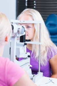
Comprehensive Eye Exams for Vision Assessment
We recommend routine eye exams. But, most visits to the eye doctor are to adjust prescriptions for patients that are not seeing well. The process of determining your prescription is call the refraction. While the refraction is only a small part of the tests that we will perform during your eye exam, the accuracy of those results determines how happy you will be with your new correction.
Other tests performed during your routine eye exam are:
• Slit lamp examination of the structures in the front of the eye, including eyelids, conjunctiva, sclera, lens, iris, and cornea
• Examination of the structures in the back of the eye, including the retina and optic nerve
• Refraction to determine vision correction needs
• Cataract screening
• Glaucoma screening and internal eye pressure checks
• Corneal and retinal evaluations
• Visual field examination
Medical Eye Exams
Your eyes rarely hurt when there is something wrong. Many eye diseases can be detected through routine eye exams and treated effectively before they cause damage that cannot otherwise be repaired. In addition, a variety of whole-body diseases can be identified during an eye exam and be treated before they cause permanent damage. That is why we include eye health assessments in our routine eye exams for vision.
If you experience sudden changes in vision or unusual visual symptoms you should call to schedule an immediate visit. If we diagnosis eye disease our relationship with world-renowned ophthalmologists ensure that our patients receive the best overall medical and visual care.
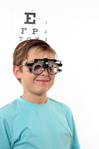
Eye Exams for Children
Most pediatricians are trained to monitor the development of your child’s eyes and vision. They will advise you of the need for early intervention by a pediatric ophthalmologist if it is indicated. These specialists’ can use modern techniques to accurately exam the eyes of even the youngest patients.
Normally, your child’s first comprehensive eye examination should coincide with their ability to recognize letters. This is usually around the time your child enters the first grade. At this visit the we will evaluate your child’s eye health, the coordination of the muscles that control eye movements and measure for a prescription to convert their vision needs.
We are also pleased to provide the following services:
• Preoperative and postoperative LASIK consults
• Corneal reshaping using contact lenses
• Ocular allergy therapy
• Dry eye therapy, including punctal plugs and tear film assessment
HMO’s and Insurance Plans:
We participate in and provides services for most HMO’s and Insurance plans. Vision benefits vary widely. Most health insurances offer eye exams with a small copayment. Some plans also offer direct reimbursements for your eyeglass or contact lens purchase. Other programs allow you to pick products from specific collections. More commonly, Parrelli Optical® offers a substantial savings in the form of a discount taken off the list price of eyewear for our patients who are covered by various health insurance programs.
Specific information about your insurance plans can be found at your insurer’s website:
Aetna BlueCross/BlueShield of MA Eyemed Harvard Pilgrim Health Plan MassHealth Medicare Neighborhood Health Plan Network Health Tufts Health Plan Tufts Medicare Preferred Unicare/GIC United Health Care
Or you can call us for specific information about your coverage. Have your enrollment information handy and we’ll do the rest.